Choroidal metastasis


A diabetic 55 years old lady with a history of breast cancer presented to me with visual loss in her left eye.


Fundus image 


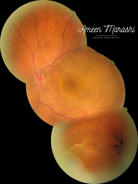
Her fundus exam shows chorioretinal elevation in the superior temporal quadrant with pigmentary changes and intraretinal blood. In contrast, the left eye examination shows multifocal retinal serous detachments at the macula and inferior retinal quadrants.


Fundus image of the Right Eye sub retinal fluids from metastatic choroidal mass
Fundus image of the Right Eye sub retinal fluids from metastatic choroidal mass 

Fundus image of the Left Eye showing RD from exudative metastatic choroidal mass
Fundus image of the Left Eye showing RD from exudative metastatic choroidal mass 

Optical coherence tomography


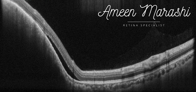
OCT of the right eye shows an elevation of the chorioretinal complex with subretinal fluids due to metastatic mass. In contrast, the left eye OCT scan shows an elevation of a choroidal retinal complex with massive subretinal fluids and some intraretinal cysts.
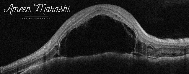
OCT cross-section showing choroidal mass altering chorioretinal contour with subretinal fluids

OCT cross-section showing choroidal mass altering chorioretinal contour with subretinal fluids


Diagnosis is choroidal metastasis in the right eye while the left eye is serous retinal detachment due to choroidal metastasis.


Differential Diagnosis


-Choroidal amelanotic melanoma -Posterior scleritis -Choroidal osteoma

-Harada

-Central Serous Retinopathy


Management


Systemic chemotherapy along with plaque radiculopathy


Discussion


This lady was diagnosed with breast cancer and managed with mastectomy and hasn't been followed up for years. Her diabetes has nothing to do with current pathology and detachments. This case shouldn't be confused with amelanotic melanoma as metastasis due to a history of primary breast cancer and presented as unifocal and pigmentary changes. At the same time, Posterior scleritis features pain and choroidal folds, which are absent in this case.


This case is easily differentiated from central serous retinopathy due to general thin choroidal profile (in except area of the choroidal mass area) and the intraretinal cystic formation, and finally, this cannot be mistaken with Harada due to the natural history of the patient and elevation of the choroidal retinal complex is the prominent feature rather than multifocal serous retinal detachments.


Management usually should be done with chemotherapy under an oncologist's supervision, and pars plana vitrectomy is NOT indicated.

Comments

  1. You have provided valuable data for us. It is great and informative for everyone. Keep posting always. I am very thankful to you. eye specialist in Houston

    ReplyDelete

Post a Comment

Popular posts from this blog

Cellophane maculopathy

Caused by an epiretinal membrane, clinically, it appears as a sheen abnormal foveal reflex which features epiretinal membrane, due to inflammatory mediators and proliferation of glial cells secondary to a partial separation of posterior cortical vitreous. Fundus image shows cellophane maculopathy OCT features a hyperreflective band covering the inner retinal layers, causing minimal disturbance of inner retinal tissues. OCT cross-section shows epiretinal membrane  Cellophane maculopathy itself in individuals with good visual acuity may not need any interventions, but it is warranted to do a detailed peripheral retinal exam to rule out retinal tear or hole However, an epiretinal membrane may proliferate further and cause additional tangential traction. Thus, it may induce macular pucker or lamellar/pseudo hole and increase disturbance of inner retinal tissues, macular thickness, and reduction of vision. These patients may require pars plana vitrectomy to remove the epiretinal membrane an

RPE tear, and it's OCT features in a nutshell

RPE tear can occur in vascularized PEDs due to a complication of intravitreal AntiVEGF treatment, which causes a rapid contraction of CNVm, inducing traction on the RPE surface and shrinking of RPE surface. On OCT, the RPE rip will indent the retina with increased hyperreflectivity in contracted RPE with underlying shadowing hinders choroidal view in contrast to the area where RPE where it ripped of which only Bruch's membrane is visible and increasing the visibility of the underlying choroid. RPE tear Thus two opposite forces, the regression of CNVm and remaining attached RPE, other causes such as laser or spontaneous can occur. However, the AntiVEGF treatment continued despite the RPE tear. Please check  Clinical guidelines for ARMD  and  course of clinical changes in RPE

Sectoral retinitis pigmentosa

Sectoral retinitis pigmentosa is an autosomal dominant and usually associated with acceptable visual acuity.  The fundus examination shows pigment clumps and chorioretinal atrophy only in sectoral with almost normal optic disc configuration and vessel diameter. Fundus image showing Sectoral retinitis pigmentosa The sectoral retinitis pigmentosa symmetrical in both eyes Perimetry will show scotoma corresponding to the dispersion of pigmentary clumps. OCT shows outer retinal degeneration and RPE thining in the area of pigmentary clumps. Please check  Clinical guidelines in retinitis pigmentosa