Would you consider this case of early PDR associated with diabetic macular edema?

A 58 years old male diabetic for ten years with BCVA 20/20 presented to me for a regular eye exam with IOP 12 mmHg.


Fundus Image


His Fundus photograph shows hard exudates far from the fovea with microaneurysms, intraretinal dot blood, and soft exudates (cotton wool spots) where the optic disc has a deep configuration (bucket like) with asymmetric orange to red color.
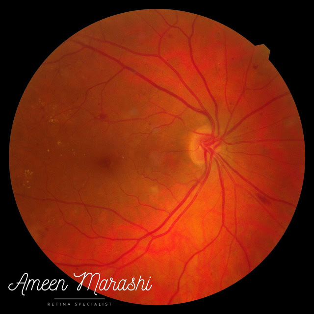


Fundus image of subtle NVD
Fundus image of subtle NVD 


Optical Coherence Tomography 


OCT scan shows normal foveal contours and thickness with the absence of diabetic macular edema.

Normal OCT scan despite leaking microaneurysms


Fundus fluorescein angiography 


FFA shows hypofluorescence in the mid retinal periphery due to hypoperfusion (ischemia) with hyperfluorescence dots and hyperfluorescence superior temporal of the optic disc; this hyperfluorescence increases and develops a leak in the mid and late stages.


Early phase of FFA showing leakage from the optic disc


Mid phase of FFA showing leakage from the optic disc


Late phase of FFA showing leakage from the optic disc


Diagnosis


Early PDR with NO diabetic macular edema


Management


Pan Retinal Laser photocoagulation (PRP) in 2 sessions two weeks apart


Discussion


This eye has reached the state of relative ischemia, as shown in FFA in the form of peripheral hypofluorescence. Despite the leaking microaneurysms in the macula, there is no diabetic macular edema. 


However, the early hyperfluorescence and leakage from the optic disc show small neovascularization (NVD) that was subtle in fundus photography. NVD is small in size with a bucket configuration of the optic disc.


The PRP was chosen as mainstay treatment in two sessions two weeks apart as there is no diabetic macular edema and not to induce macular edema. However, OCT follow-up is mandatory.


Take-home messages


1) When we see hard exudates or leaking microaneurysms on FFA, that does not mean there is diabetic macular edema

2) OCT is the gold standard to diagnose diabetic macular edema

3) FFA can help us diagnose ischemic changes and subtle neovascularization even when the fundus looks quite like this case

Please check clinical guidelines in diabetic retinopathy

#OCT #FFA #PDR #PRP #Diabetic #Laser #DME #Leakage #NVD #Neovascularization


Comments

Popular posts from this blog

Adult-onset vitelliform macular dystrophy

One of the most common pattern dystrophies similar to best occurs in adulthood, as it appears as oval or yellowish macular material, originated from the debris of photoreceptors, or the RPE phagocytosis failure. Fundus image showing Adult-onset vitelliform macular dystrophy  On OCT, it appears as subretinal vitelliform material accumulation but above the RPE with intact ellipsoid zone and thus explains good visual acuity without an increase in retinal thickness. OCT cross-section showing Adult-onset vitelliform macular dystrophy Fluorescein angiography typically shows hypo-fluorescence at the area of vitelliform accumulation surrounded with hyper-fluorescence increases intensity in late phases; however, it may show liner, patchy, or ring-like, which is inconsistent with visual acuity. Adult-onset vitelliform macular dystrophy itself does not warrant any treatment; however, a regular follow up to monitor any progression to choroidal neovascularization. How would you approa...

Cellophane maculopathy

Caused by an epiretinal membrane, clinically, it appears as a sheen abnormal foveal reflex which features epiretinal membrane, due to inflammatory mediators and proliferation of glial cells secondary to a partial separation of posterior cortical vitreous. Fundus image shows cellophane maculopathy OCT features a hyperreflective band covering the inner retinal layers, causing minimal disturbance of inner retinal tissues. OCT cross-section shows epiretinal membrane  Cellophane maculopathy itself in individuals with good visual acuity may not need any interventions, but it is warranted to do a detailed peripheral retinal exam to rule out retinal tear or hole However, an epiretinal membrane may proliferate further and cause additional tangential traction. Thus, it may induce macular pucker or lamellar/pseudo hole and increase disturbance of inner retinal tissues, macular thickness, and reduction of vision. These patients may require pars plana vitrectomy to remove the epiretinal membran...

Approaching serous PED

Serous PED is a result of fluids leak via Bruch’s membrane to space between Bruch’s and RPE, causing an increased in hydrostatic pressure separating RPE from Bruch’s membrane. Fundus image  On clinical examination, a smooth, well-defined border of RPE elevation along with a slight pale appearance PED is associated with subretinal fluid or intraretinal hemorrhage. Fundus image showing serous PED with subretinal fluids and drusen Optical coherence tomography  On OCT, PED features smooth or irregular RPE elevation, which contains clear fluids that looks like a homogeneous hyporeflective area. PED may feature sub / intraretinal fluids accompanied by ellipsoid zone disruption. OCT cross-section showing serous PED with subretinal and intraretinal fluids Fluorescein fundus angiography On FFA, serous PED shows a distinct area of early hyperfluorescence, which increases in the mid and late phase due to dye pooling. FFA is useful to detect the presence of neovascular membrane. Fundus im...