Fibrovascular PED Case report and Discussion

An 81 years old man pseudophakic presented to me with features of ARMD; his BCVA is 20/200 in his Right eye, while in his left eye, the BCVA is 20/35; fundus examination showed drusen and signs of ARMD.


Fundus image


Fundus examination shows drusen, RPE elevation, and subretinal fluid.

Fundus image shows fibrovascular PED with reticular pseudodrusen


Optical coherence tomography


OCT cross-sections show RPE detachment area induced by a fibrovascular membrane with pronounced Bruch’s membrane forming double layered sign and subretinal fluid along of disturbed ellipsoid zone.

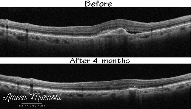
The upper OCT cross-section shows fibrovascular PED with subretinal fluids the lower OCT cross-section shows decreased size of PED and resolved subretinal fluid four months post treatment with monthly AntiVEGF
The upper OCT cross-section shows fibrovascular PED with subretinal fluids the lower OCT cross-section shows decreased size of PED and resolved subretinal fluid four months post treatment with monthly AntiVEGF 


Diagnosis: This is a case of wet ARMD with occult choroidal neovascularization.


Management: This patient managed with monthly intravitreal Aflibercept for three consecutive injections and one-month post-treatment OCT cross-section shows resolving both subretinal fluid and fibrovascular PED and improved vision to 20/50, and the patient was followed up using PRN protocol.


Discussion


Wet ARMD can present as occult Choroidal neovascularization, which features a vascular proliferation sub RPE, and this proliferation may detach the RPE and induce PED and subretinal fluid, causing disturbance of outer retinal layers (ellipsoid zone),

Subretinal fluid and occult choroidal CNVm have a better visual prognosis than classical choroidal neovascularization, and intraretinal cyst may have a worse prognosis due to subretinal scar formation and intraretinal tissue disorganization.

This case treated with intravitreal Aflibercept for three consecutive monthly injections and one-month post-treatment OCT shows improvement in PED regression showing regression of the fibrovascular membrane and reducing in size and resolved subretinal fluid with BCVA improved to 20/50 and not better due to the disturbed ellipsoid zone.


Please tell me how you would manage this case in your clinical practice??!!!
Please check Clinical guidelines for ARMD and course of clinical changes in RPE

Comments

Popular posts from this blog

Cellophane maculopathy

Caused by an epiretinal membrane, clinically, it appears as a sheen abnormal foveal reflex which features epiretinal membrane, due to inflammatory mediators and proliferation of glial cells secondary to a partial separation of posterior cortical vitreous. Fundus image shows cellophane maculopathy OCT features a hyperreflective band covering the inner retinal layers, causing minimal disturbance of inner retinal tissues. OCT cross-section shows epiretinal membrane  Cellophane maculopathy itself in individuals with good visual acuity may not need any interventions, but it is warranted to do a detailed peripheral retinal exam to rule out retinal tear or hole However, an epiretinal membrane may proliferate further and cause additional tangential traction. Thus, it may induce macular pucker or lamellar/pseudo hole and increase disturbance of inner retinal tissues, macular thickness, and reduction of vision. These patients may require pars plana vitrectomy to remove the epiretinal membran...

RPE tear, and it's OCT features in a nutshell

RPE tear can occur in vascularized PEDs due to a complication of intravitreal AntiVEGF treatment, which causes a rapid contraction of CNVm, inducing traction on the RPE surface and shrinking of RPE surface. On OCT, the RPE rip will indent the retina with increased hyperreflectivity in contracted RPE with underlying shadowing hinders choroidal view in contrast to the area where RPE where it ripped of which only Bruch's membrane is visible and increasing the visibility of the underlying choroid. RPE tear Thus two opposite forces, the regression of CNVm and remaining attached RPE, other causes such as laser or spontaneous can occur. However, the AntiVEGF treatment continued despite the RPE tear. Please check  Clinical guidelines for ARMD  and  course of clinical changes in RPE

Diabetic macular edema with ischemic changes associated with very severe NPDR

A 51 years old male known to be diabetic for ten years with excellent glycemic control profile along with normal blood pressure presented with BCVA 20/100 in his left eye; he had a better vision of 20/30 in the same eye one year ago when he used to be on intravitreal injections of aflibercept, but he decided to discontinue intravitreal injection as been advised by another physician. Fundus image His fundus image shows cystic formation at the fovea with a sign of intraretinal dot and blots hemorrhages in the mid periphery with cotton wool spots and IRMA (Intra Retinal Microvascular Abnormalities). Fundus image showing macular edema with intraretinal hemorrhage and cotton wool spots Optical coherence tomography His OCT scan shows thickened central macular tissues with disorganization of inner retinal layers and non-empty cystic formation as it is classic findings of diabetic macular edema with some empty cystic formation; however, the ellipsoid zone looks intact. OCT cross-section for ce...